
Cryo-EM structures of a bacterial efflux pump and a fungal-specific channel show how to tell low permeability apart from active expulsion of a drug candidate.
A drug candidate can pass the whole initial screening (exploratory and in vitro phase), showing the expected potency, acceptable selectivity and cytotoxicity under control. Yet it is common for the effect simply not to appear in in vivo studies.
The first hypothesis is usually permeability: the molecule is not crossing the barriers. There is, however, a second possibility, harder to prove without the right tool. The molecule does get in, but it is recognised and expelled by a membrane transporter before it can act. Solving this is complex, because one calls for chemical reformulation of the molecule and the other for a co-administered efflux inhibitor, or even a complete change of target.
For decades, answering this question precisely while still in the in vitro phase was practically impossible. Membrane transporters and channels are hard to analyse by X-ray crystallography, long the most reliable way to see a protein in atomic detail. Membrane proteins rarely form the ordered crystals the technique requires and tend to destabilise during purification. Without a structure, only inference remains: indirect models, comparison with homologous proteins and bench trial and error.
Seeing what could only be assumed
Cryo-electron microscopy changes this equation. The sample is frozen in fractions of a millisecond, in a state and conformation close to what exists inside the living cell, and the three-dimensional structure is reconstructed from thousands of individual particle images captured by an electron microscope. Nothing needs to be crystallised. When resolution reaches the 2 to 3 ångström range, the result makes it possible to locate, atom by atom, the path a molecule takes inside a channel, the exact point where it binds and the movement that opens or closes the passage. Two recent examples, one in a bacterium and one in a fungus, published between late 2025 and early 2026, show the reach of this.
TMexCD1-TOprJ1, tigecycline efflux pump in Klebsiella pneumoniae
TOK1, fungal-specific potassium channel, in Candida albicans
The pump that refuses to let go of the antibiotic
In November 2025, a group of researchers published in Nature Communications the cryo-EM structure of TMexCD1-TOprJ1, a tripartite efflux pump originally identified in Klebsiella pneumoniae. The pump actively expels tigecycline, an antibiotic reserved as a last resort against multidrug-resistant infections, and the problem is not restricted to this bacterium: the gene encoding the machinery lives on mobile plasmids that can circulate among different ESKAPE pathogens, the bacteria of greatest concern to global health surveillance. When this resistance appears alongside other mechanisms, such as the MCR-1 gene associated with colistin resistance, the combination further narrows the available therapeutic options.
The structure, solved at 2.97 ångströns, revealed an unusual symmetric architecture, with three copies of TOprJ1 in the outer membrane, six of TMexC1 and three of TMexD1, forming an elongated assembly that spans both bacterial membranes like a continuous duct. The tip-to-tip contact between the TMexC1 adaptor and the TOprJ1 pore, and the cavity where the antibiotic binds before being pumped out, are now mapped precisely enough to guide the design of molecules that specifically block this interface without interfering with other pumps in the cell.
A channel with no human counterpart
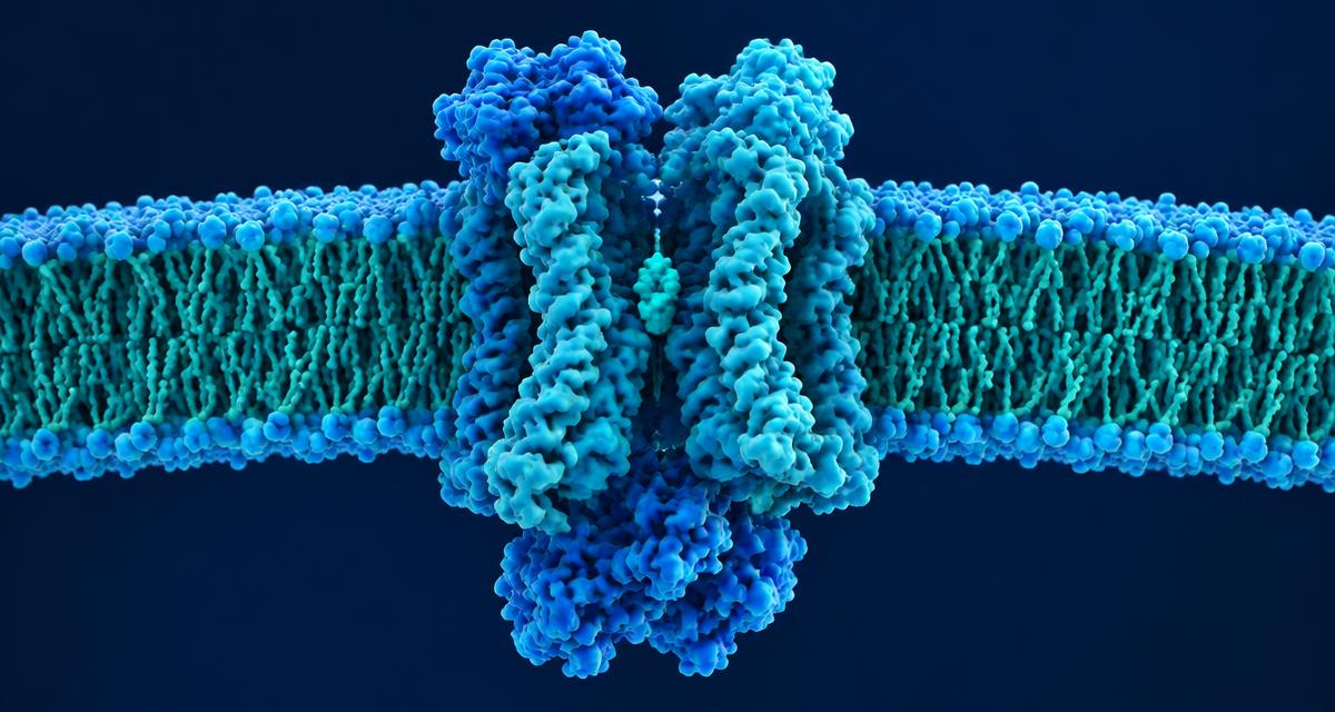
The second example is of a different nature but follows the same logic. In April 2026, Nature Communications published the structure of TOK1, a fungal-specific potassium channel found in Candida albicans, the most common fungal pathogen in human infections. TOK1 regulates the fungal cell's potassium balance under stress, sustaining the internal pressure the cell needs to colonise the host, and has no clear homologue among human potassium channels.
The structure, obtained at 2.6 ångströns, showed eight transmembrane helices arranged differently from known mammalian channels, a domain named TALC that acts as an external pH sensor and resembles auxiliary subunits found in neurons, and an opening mechanism that depends on a helical movement locked at a single amino acid, alanine 471. Potassium selectivity also stood out: instead of the four classic coordination sites present in most channels of this type, TOK1 uses only three. It is precisely this kind of structural difference, distant from human biology, that interests those looking for a new antifungal target with lower risk of side effects on human ion channels.
What changes in practice for developers
None of these structures, on its own, solves a drug development problem. Their value lies in bringing together two dimensions that do not always talk to each other: the behaviour observed in biological assays and the molecular mechanisms that explain it.
In practice, this means apparently contradictory results can be investigated with more specific hypotheses. If a candidate shows good in vitro activity but loses efficacy in more complex models, for example, the investigation can move beyond the low-permeability hypothesis. Identifying efflux transporters, binding sites and conformational changes helps target functional assays to check whether the molecule is being recognised and expelled by the cell. Likewise, structural knowledge of channels and transporters unique to certain pathogens opens possibilities for designing more selective compounds.
For preclinical development teams, the impact is on the quality of decisions made throughout the research: choosing which mechanisms to investigate, which assays to prioritise and which structural changes may increase a candidate's efficacy or selectivity. Cryo-EM does not replace functional experimentation, but it adds a layer of information that makes the investigation more targeted and shrinks the space for trial and error.